Categories
EEG Swartz Center/UC San Diego

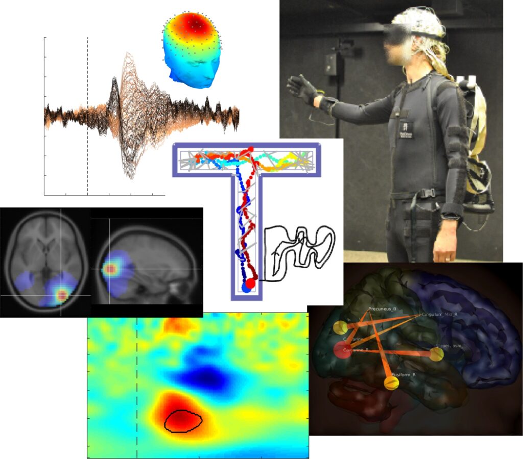
Publication: The Audiomaze: An EEG and motion capture study of human spatial navigation in sparse augmented reality

Journal page

https://onlinelibrary.wiley.com/doi/epdf/10.1111/ejn.15131

AudioMaze is Swartz Center’s main Mobile Brain-Body Imaging (MoBI) projects that was multi-million-dollar funded, and the dedicated lab has been in ‘the downstairs’ for more than a decade. Basically, the idea is that a blindfolded subject walks through the digitally defined mazes which has in programmable walls that returns audio feedback when touched, hence AudioMaze. Because blindfolded subjects need to walk very slowly, and the spatial probing is limited to stretching the right arm, subject’s behaviors becomes very slow, stereotypical, and repeated, all of which is suitable for event-related brain potential analysis.

The highlight of the finding is that the occipital regions, which is typically associated with visual processes, showed modulated activities although the subjects were blindfolded. My colleagues in Berlin has already published dozens of navigation related studies and suggested that retrosplenial cortex, which is right under the posterior cingulate cortex, is involved in this process. Generally speaking, that could be the truth of functional brain mapping. However, when it comes to detecting retrosplenial cortex with scalp EEG recording, it sounds too ambitious for me. I found in literature that lingual gyrus is a posterior part of parahippocampal place area (PPA), which seems to fit as the explanation of the result I got from the study. So I put more weights on the lingual gyrus in the interpretation.

As an experimental approach in EEG analysis, I used group-level source information flow toolbox, groupSIFT, to calculate effective connectivity across probabilistic independent component densities resolved in the brain regions at the group-level analysis. Not surprisingly, I found time-evolving effective connectivity networks centered at the occipital and inferior parietal regions.

This paper has 25 pages, 12000 words, and 14 figures (plus 3 additional figures in the Supplement). I told grad students and post-docs that they should not write a paper like this because it risks their careers. The experimental paradigm is new, the analysis is new, no psychologically clever top-down question is available, etc. In a sense, it was good that I took this job because I was old enough.

Categories
EEG Swartz Center/UC San Diego

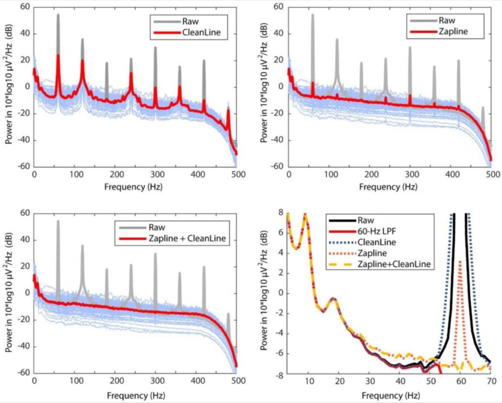
Publication: Can We Push the “Quasi-Perfect Artifact Rejection” Even Closer to Perfection?

This short technical paper was originally written to an opinion paper to ZapLine paper.

https://www.sciencedirect.com/science/article/pii/S1053811919309474

Therefore, the title makes sense only in that context. However, because the submission was rejected, now it does not make much sense. One of the coauthors pointed it out to me only after publishing the paper.

Journal page

https://www.ncbi.nlm.nih.gov/pmc/articles/PMC7873913/

The point of this paper is that even though ZapLine is an amazing line-noise reduction solution by itself, as the original author called it ‘quasi-perfect’ in the highlight points, there is actually an easy way to improve the performance. That is, to apply CleanLine after ZapLine. Why can I predict it? It is because ZapLine is a spatial filter method, while CleanLine is a non-stationary method in the time domain.

One of the reviewers said this is not worth a paper because there is no novel finding or development. Yes, it has a good point. But at the same time I believe that sharing this idea supported by an insight and evidence is also an important part of scientific communication.

By the way, the original author of ZapLine, Alain de Cheveigné kindly helped me to write this paper, although I was teasing his phrase ‘quasi perfect’ in the manuscript. I deeply appreciate his generousity!

Categories
EEG Swartz Center/UC San Diego

Publication: Cross-Frequency Power-Power Coupling Analysis: A Useful Cross-Frequency Measure to Classify ICA-Decomposed EEG (PowPowCAT reference paper)

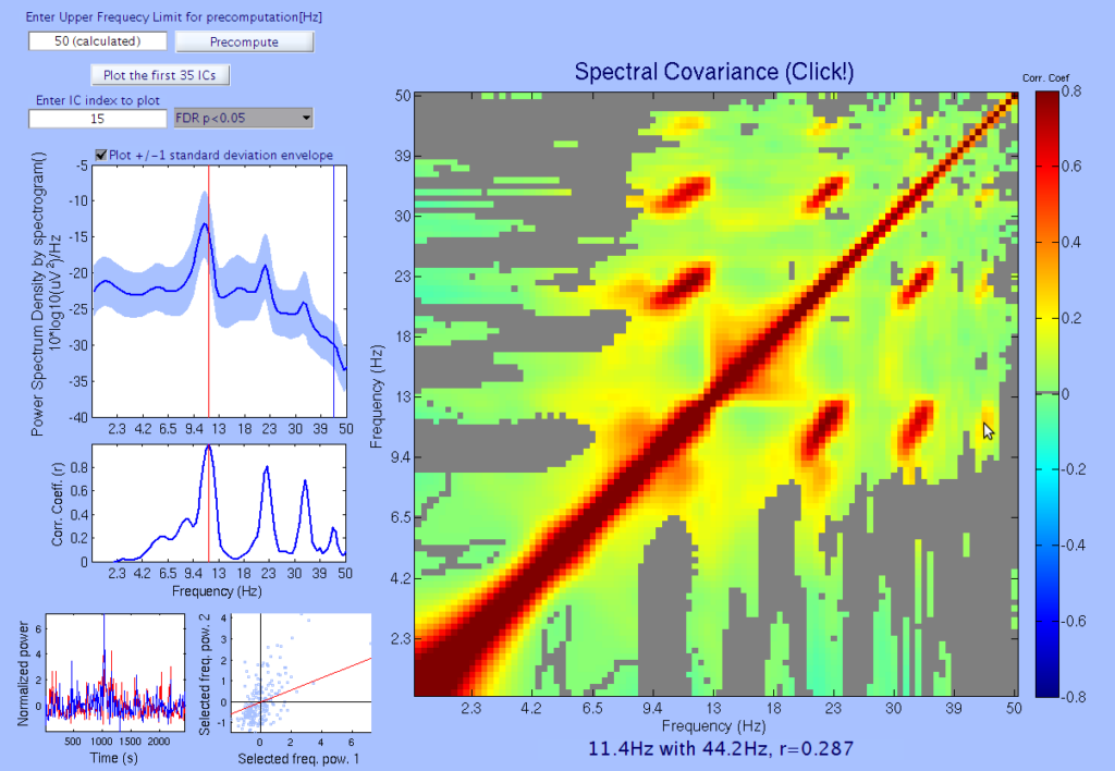
The first author Nattapong visited me at the Swartz Center in UCSD a few years ago. At that time, I was fascinated by re-inventing (without knowing, of course) the idea of comodugram/comodulogram, which is a visualization of cross-frequency power-power coupling (also called spectral covariance). I suggested the publication of this idea to Nattapong, and he agreed. Then the idea was published.

Journal page

https://www.mdpi.com/1424-8220/20/24/7040/htm

EEGLAB plugin Wiki page

https://github.com/sccn/PowPowCAT

Sometimes you see two peaks in power spectral density (PSD) of EEG signals. But have you seen four peaks!? The owner of this brain is a grad student in our lab.

This comodulogram has been probably most frequently used in Buzsáki lab in the field that is closest to mine. In fact, Nattapong and I wrote them to ask about it. They were so kind, György pointed us to several of his colleagues and they told us several interesting things about this method.

Initially, I did not even know the name of this visualization. I asked it to a couple of smart people, but no one knew it. Later, Nattapong and I learned that it is called ‘comodugram’. But I did not like the name. The other, slightly less common name ‘comodulogram’ sounds better. But I like the fully descriptive version of the name, Power-Power Coupling Analysis Tool (PowPowCAT). Actually, I would be motivated to publish any computational tool that has the name as nice as PowPowCAT.

Categories
EEG Swartz Center/UC San Diego

Publication: Modulation of Frontal Oscillatory Power during Blink Suppression in Children: Effects of Premonitory Urge and Reward

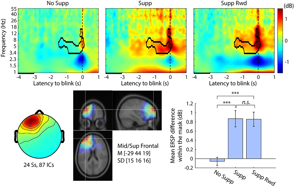
This is a part of a study on chronic tic disorder conducted by Sandra Loo at UCLA. The idea is to investigate the effect of blink suppression in typically developed children. She included a unique idea of instructing the children to use ‘urgeometer’, which is just a customized joystick, to self-report the amount of urge in real time. Basically, I could find a pretty straightforward prefrontal involvement during voluntary blink suppression.

The link to the full paper (open access) is here.

https://academic.oup.com/cercorcomms/article/1/1/tgaa046/5881803

There was actually another intention in this paper, which is more related to EEG methodology. Think about the reason why this kind of study has not been done so far–it is because eye blink is a catastrophic artifact for a standard EEG analysis. I thought it is my responsibility to show minimum level of validation to justify my blink-related EEG analysis. This endeavor is, not surprisingly, reported in the Supplement.

https://oup.silverchair-cdn.com/oup/backfile/Content_public/Journal/cercorcomms/1/1/10.1093_texcom_tgaa046/1/supplementary_material_revision1_tgaa046.docx?Expires=1616171516&Signature=syI9xbw3xWlhjqmNZVhLRacw2pwi8CjXFQhWyo8fiertNMOdIFh80IyS6UZTs3QCtANgEFTfu89sFJpi1lexyNuLX-UGdJzEKWWbAqp0DwW3oEHh0EapvQYalfj33BnqZz5GglE0LBMde-L7VU~CZl63KqWd4Ilg1YY5rq7kV3p735YUGShpnraU8KSmW6qzsbzUpVhrJYoQnuyT-uBjS~-dzql476zQI5xobte3WiOkciUPPsQBYv9YdmJ8KeuKQZ~vBTut43pqjgq3hcj4OT4Se2gEoN2x63ayw3VwiFjUwONiM7f2u2fBxg~M7MYXY56lx6xMZgHeMJKJw-KCeQ__&Key-Pair-Id=APKAIE5G5CRDK6RD3PGA

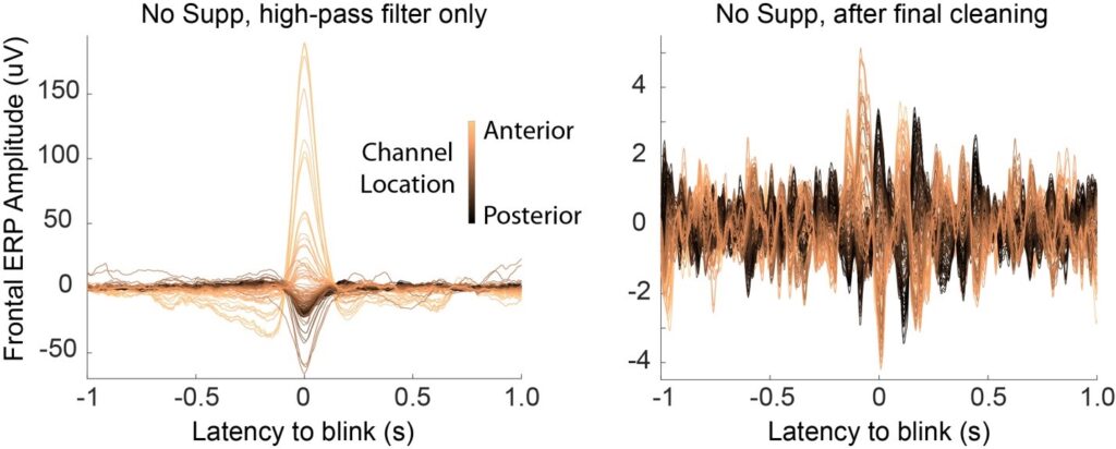
For example, this is the before-after comparison. Apparently, the amplitude scale is 40 times different in this comparison! According to what I reported, average 98.4% of signal variance was removed from scalp-electrode signals. I thought this number is insane, so I wanted to check it.

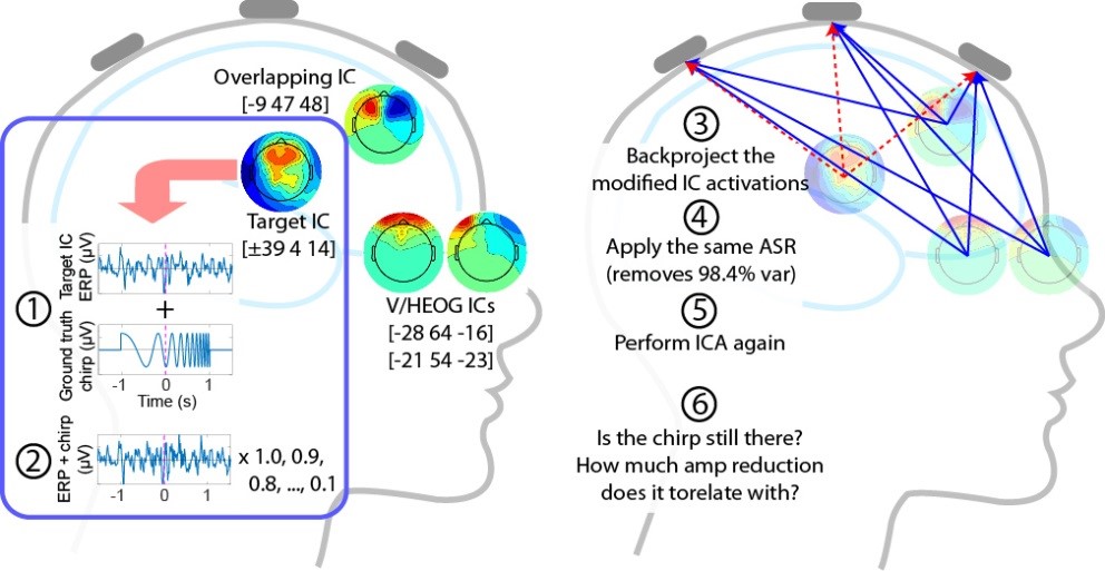
How can I show that bathwater was thrown out, not the baby? The right way to do it would require a well-designed simulation study, which I could not afford. So I attempted to show the best circumstantial evidence that is available as a post-hoc validation using an ok-level-designed simulation.

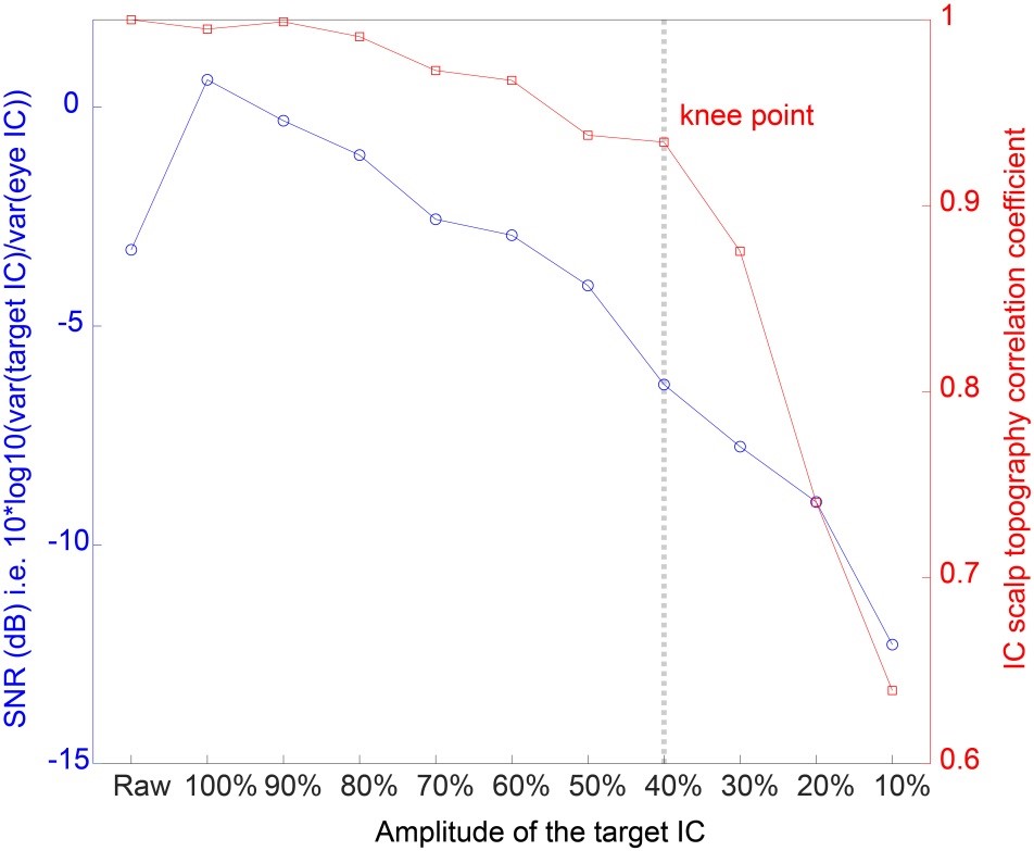
I added a small ground-truth signal (1% of unprocessed signal standard deviation) in the scalp signal, then applied the data cleaning method, artifact subspace reconstruction (ASR) + independent component analysis (ICA) to see if the ground-truth signal survives. This process was repeated for the different levels of SNR which was progressively lowered. The result was like this.

I found that the IC-scalp topography became suddenly worse when SNR is lowered to less than 30%. So I concluded that the small signal (1% of unprocessed signal standard deviation) can easily survive the current ASR + ICA methold. Moreover, it has further SNR margin of 7dB.

This ok-level-design was whipped up for a quick proof of concept, but it seems to deserve further development into a full method paper. However, I have so many of this kind of ideas that I can’t publish all of them. If there are grad students or post-docs interested in this idea, I would be happy to collaborate.

Categories
EEG GYREE LAB Swartz Center/UC San Diego

Ground truth of EMG removal: Paralysis vs. ASR + ICA

For automated scalp-EEG denoising, I always use EEGLAB plugin clean_rawdata(), which includes now popular artifact subspace reconstruction (ASR), and ICA. The clean_rawdata() was written by my former colleague Christian Kothe as a offline-version of his BCILAB data cleaning pipeline upon my request. I just wrote a wrapper for it to make it into an EEGLAB plugin. I reported this historical fact in the Section 6 of this Supplementary Materials for record https://academic.oup.com/cercorcomms/article/1/1/tgaa046/5881803#207580614 ASR is an ideal preprocessing for ICA due to compensatory properties. ASR uses non-stationary approach (sliding windows), while ICA uses stationary approach (a single spatial filter that is suppose to be valid for every data point). ASR can handle artifacts with an astronomically large outlier, which can cause instant death to ICA, or even if does not kill it will certainly destroy the result into nonsense (Have you ever seen explosive scalp topos?) Controlling the performance of ASR is not as easy as applying ICA (which is just a button press), but the rule of thumb is that use SD = 20 and you are fine (for quantitative evidence, see my colleague Chiyuan’s paper https://ieeexplore.ieee.org/abstract/document/8768041…Conclusions: Empirical results show that the optimal ASR parameter is between 20 and 30, balancing between removing non-brain signals and retaining brain activities.“)

However, this advocation of the performance of ASR + ICA is only qualitative. How can the performance be proven? Here comes in the eternal-recurrence problem of EEG research: the lack of the ground truth. EEG research is doomed by this problem. The vacancy of the ground truth corroded EEG researcher’s mind so that now they believe more in colorful figures produced by fancy-sounding signal processing techniques than scientific thinking. Once I pointed out that it is an erroneous attempt to compensate science with engineering–we should ask ourselves if we are using the colorful figures to stay closer to the ground truth. Fortunately, someone knowledgeable told me about a nice study that shows ground truth of EMG contribution to scalp-recorded EEG (Whitham et al., 2007 https://www.sciencedirect.com/science/article/abs/pii/S1388245707001988?via%3Dihub). I like this study so much that this time I decided to pay $48.30 to Elsevier to get the license to show their nice plot for you.

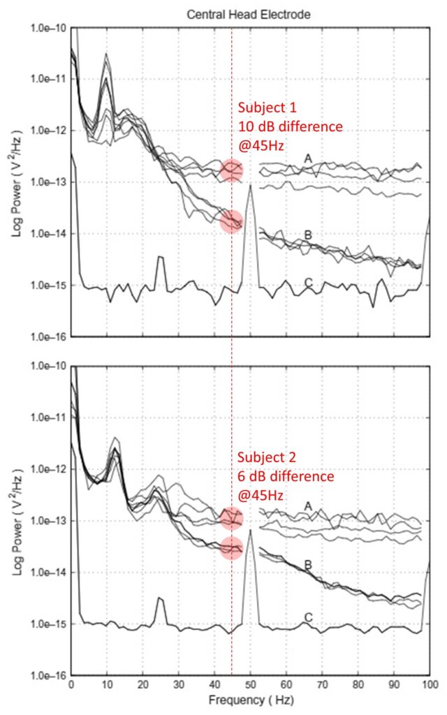
Whitham et al. (2007) Clinical Neurophysiology 118:1877–1888 Figure 1. Replicated with permission. PSD (n=2) calculated at the electrode between CPz and Pz (linked-ear reference). A, without paralysis. B, with paralysis (20 mg of cisatracurium by intravenous injection). Muscular paralysis was evaluated by right common peroneal nerve stimulation to extensor digitorum brevis. The motor action potential was confirmed to be disappeared in 5 min after the administration.

You see a ordinary-looking PSD plots from two subjects in the top and the bottom plots, but there are curves A and curves B. What do you think is the difference between A and B in each plot? The answer is muscular paralysis. Because the paralyzed muscle generates zero EMG, PSD in B serves as a ground truth of EEG with no EMG. At 45 Hz, the difference between A and B are 6 and 10 dB for each subject. Let’s compare these values to our typical application of ASR + ICA.

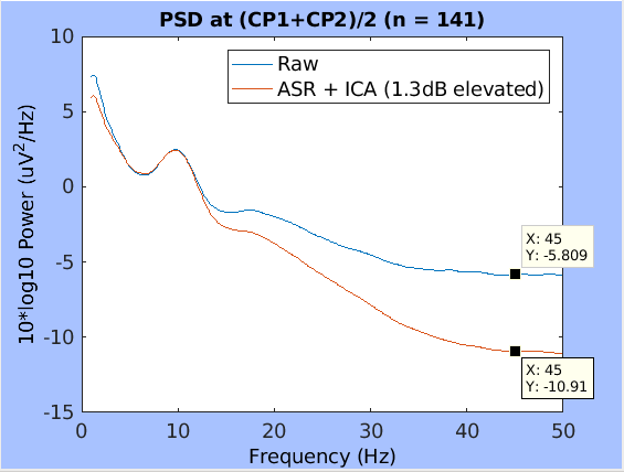
Unpublished data. PSD (n=141) calculated at the electrode CP1 and CP2 then averaged (T7-T8 average reference). Blue, raw EEG (high-pass fitler only). Red, ASR (SD=20) and IC selections (rejecting all non-brain ICs labeled by ICLabel). Note that PSD for ASR + ICA is 1.3 dB elevated to align the alpha peak power.

This plot was generated for writing a rebuttal to one of the comments made by a reviewer said ‘the performance of ASR + ICA is questionable.’ Challenge accepted automatically. There are minor differences due to different experimental settings (the same electrode location was not available, so was the initial reference) but it does not seem critical. At 45 Hz, the PSD difference between raw and ASR + ICA is about 5 dB after aligning the alpha peaks. Well, the result is not too bad.

Summary and conclusion from this comparison: (1) Using paralysis allows evaluation of the grand truth of EMG contribution to PSD of scalp-recorded EEG, which measured 6-10dB at 45 Hz; (2) ASR + ICA reduced PSD power similarly, which measured 5 dB at 45 Hz. I conclude ASR + ICA showed a good performance in comparison with the ground-truth data of EMG removal. If ASR + ICA were a simple frequency filter to mechanically suppress the gamma-band rage, it would have been trivial. However, what ICA actually does is to find a spatial filter that minimizes mutual information to achieves instantaneous temporal independence across components. So ICA is not even similar to a simple frequency filter. What we see here instead is that optimizing a mathematical property of the data reveals physiologically valid results. A similar structure is found in the argument about the origin of dipolarity of ICA-derived scalp topographies, which I once called independence-dipolarity identity (IDID). I will visit this topic some day later.

Categories
EEG Swartz Center/UC San Diego

‘Electric Field of the Brain’ Chapter 8 translated

I have been translating the second edition of ‘Electric Field of the Brain’ by Nunez and Srinivasan (2006) into Japanese since November 2018. I have not found a publisher in Japan but it does not matter. It takes 3-4 month for me to translate one chapter which is about 50 pages. There are 11 chapters in 530 pages, 12 appendices in 74 pages, and 6 pages of index list, total of 610 pages. I skipped Chapter 3 to finish an abridged version of the translation first, which was suggested by one of the publishers I talked to. The senior author Dr. Paul Nunez suggested the Chapter 3 may be omitted. I am reading while translating, so probably I am the slowest reader of this book ever. But totally enjoy the process.

I found the 1st edition of this book in SCCN’s library in the early 2010’s. I opened random pages to scan. One of the figures attracted my attention.

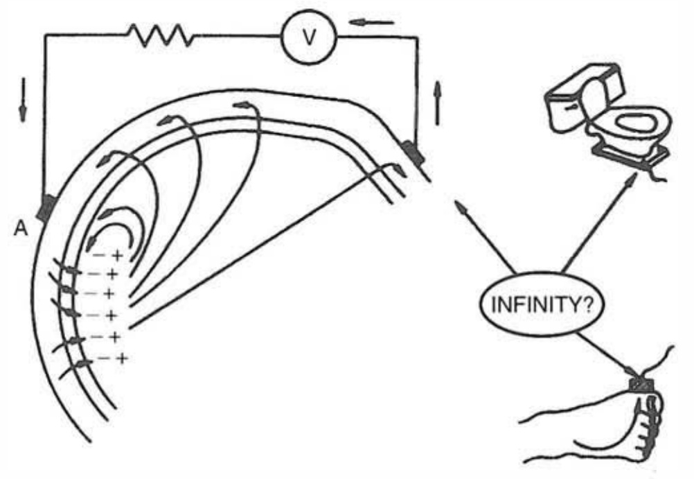
Fig 1-21 of Electric Field of the Brain (Nunez and Srinivasan, 2006) Copyright © 2006 by Oxford University Press, Inc.

On the left hand side, there are the brain, an occipital superficial dipole sheet, the resultant electric field, scalp electrodes, and a voltage meter circuit. So far so good. On the right hand space, infinity, a foot, and a STOOL. This is a graffiti, but I like the fact that the author made the authoritative Oxford University Press publish it. The figure rocks.

Another my favorite part of the book is the quote from his mentor Reginald Bickford, taken from from the Foreword to the 1st edition: ” … the authors have fallen so naturally into the lingo of the specialty while feeling free to slaughter many sacred cows that clutter the field.” This Foreword rocks too!

Categories
EEG posterPresentation Swartz Center/UC San Diego

American Epilepsy Society 2020

I made a poster presentation at American Epilepsy Society (AES) 2020. https://meeting.aesnet.org/

Automated pipeline for preprocessing scalp-recorded EEG data for phase-amplitude coupling analysis of children with and without infantile spasms
Sunday December 6, 2020
5:15 PM – 6:45 PM
Your Role: First Author

It was my first time to make a presentation in AES. It is an ongoing collaboration with neurologists in University of California Los Angeles. I worked as an analyst. My presentation was about the preprocessing pipeline.

I found a curious result that did not fit my prediction. I compared the three EEG data cleaning approaches: ASR, ASR+ICA-level1, and ASR+ICA-level2 (Result E). I found that somehow cleaning the data increased gamma power at the scalp electrodes. I should revisit this issue.